Keratoconus: early signs most people ignore — and why catching it early changes everything
Keratoconus is often missed in its early stages — but catching it early changes your treatment options significantly. Learn the signs and how Ojos Del Mar diagnoses and manages it in Tamarindo.
Keratoconus: early signs most people ignore — and why catching it early changes everything
Keratoconus doesn't announce itself dramatically. In its early stages, it looks like ordinary vision problems — blur that glasses don't quite fix, halos around lights at night, a prescription that seems to shift every time you get a new exam. By the time many patients are diagnosed, the condition has been quietly progressing for years.
Earlier detection means more treatment options. That's why knowing the signs matters.
What keratoconus is
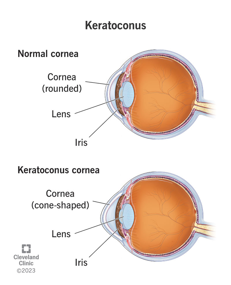
The cornea — the clear dome at the front of the eye — is normally round and smooth. In keratoconus, the corneal tissue progressively weakens and thins, causing it to bulge outward into an irregular cone shape. This distorts how light enters the eye, producing blur and distortion that standard prescription lenses can't fully correct because the problem isn't a simple refractive error — it's an irregularly shaped surface.
It typically begins in the teens or early twenties and progresses at varying rates. Some cases stabilise; others progress significantly. Early-stage keratoconus is very manageable — advanced keratoconus significantly limits treatment options.
Early signs most people attribute to something else
- Blurry or distorted vision that glasses don't fully correct — or that improves when you squint
- Halos, streaking, or ghosting around lights at night, particularly headlights
- Frequent prescription changes — needing stronger or different lenses every time you see an optometrist
- Increased sensitivity to light and glare
- Eye rubbing — keratoconus is associated with chronic eye rubbing; the rubbing itself may also accelerate progression
- One eye noticeably worse than the other
Why early detection changes your options
When keratoconus is caught early, corneal cross-linking (CXL) — a procedure that strengthens collagen bonds in the cornea — can halt progression before significant visual loss occurs. It doesn't reverse existing damage, but it stops the cornea from getting worse. This option is only effective during active progression, meaning early-stage patients who are still progressing are the right candidates.
Specialty contact lenses — including scleral lenses — can manage the vision impact even in moderate-to-advanced cases, vaulting over the irregular surface and creating a smooth optical interface. In severe cases, corneal transplant becomes the pathway to functional vision.
The difference between these outcomes is largely determined by when the diagnosis is made.
How we assess for keratoconus
Corneal topography mapping is the key diagnostic tool. It's a non-contact scan that creates a detailed three-dimensional map of the corneal surface, showing irregularities that are invisible to standard refraction testing. We include topography in comprehensive exams for any patient with unexplained vision distortion, significant astigmatism, or a history of frequent prescription changes.
If any of the symptoms above sound familiar, book a comprehensive exam or message us on WhatsApp to discuss your concerns before coming in.